The Department of Radiology at Touchette Regional Hospital provides quality diagnostic imaging services in an environment that is patient centered and customer focused. Our Board Certified Radiologists and Registered Technologists provide services to all age groups from newborn through geriatric.
Our department is divided into six clinical service areas - Diagnostic Imaging, Computed Tomography, Nuclear Medicine, MRI, Ultrasound Imaging, and Breast Imaging (Mammogram) - and our offerings include:
We are proud to offer great imaging equipment:
This equipment allows us to provide remarkable image quality, increased exam efficiency and superior care. For more information on our Radiology department or to schedule an exam, please call 618.332.5463.
Sound waves are used to visualize internal organs and structures with ultrasound. It is a fast and painless exam that does not use radiation to image the abdomen (liver, gall bladder, pancreas), OB, pelvic, breast, kidney, and vascular anatomy.
Nuclear Medicine is used to diagnose diseases of the thyroid, bone, lung, liver, gallbladder, and heart. A very small radioactive dose is either injected or swallowed, which then travels to the intended area in question. A special camera then records the activity of the radioactive dose in the form of an image. Nuclear medicine is able to monitor the function of the heart and lungs for example.
This highly technological radiology scan utilizes a series of multiple x-rays to produce detailed images of a particular area and reconstructing them as images of the internal organs and bones. Patients are often required to drink a barium solution and an injection of contrast, often referred to as dye, to help visualize internal organs and abnormalities in greater detail. CT is highly useful in scanning the brain, abdomen, pelvis, spine, sinuses, and chest.
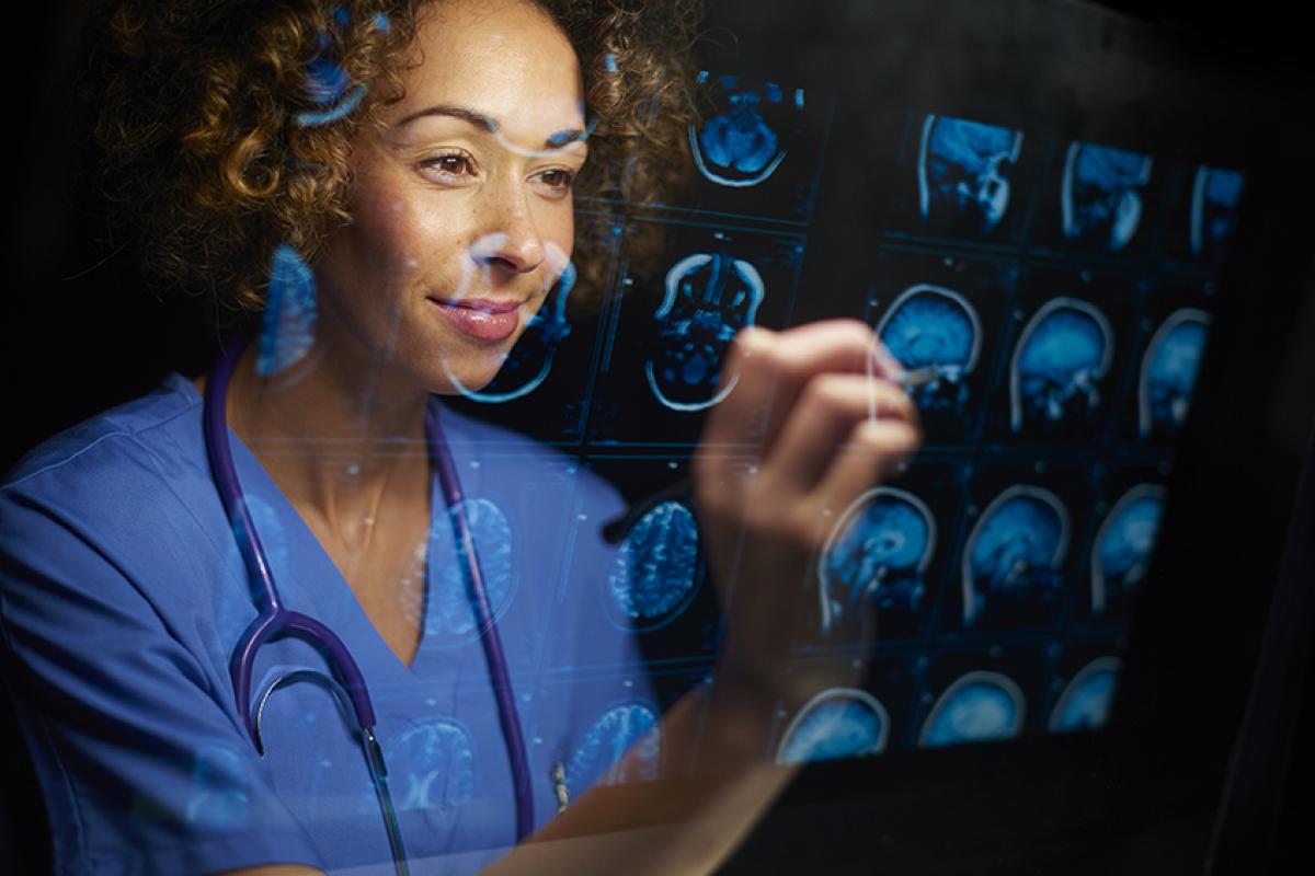
MRI uses a powerful magnet and specialized computers to visualize various part of the body without the use of radiation. The soft tissue detail demonstrated by MRI studies assists physicians to diagnose orthopedic, neurological and internal organ problems. MRI is also useful in visualizing vascular and spine anatomy.
Screening and diagnostic mammograms, as well as guided breast biopsy procedures. Our Nurse Navigator is available to assist through the process. Financial assistance is available. For more information, please call START NOW at 618.332.6130.
Radiology uses special computers to take detail images and display those images on monitors for Radiologist’s to interpret. Chest, abdomen, bones, and joints are common x-rays. Interventional radiology procedures are also available in include arthrograms with MRI or CT, lumbar punctures, myelograms are available
An Upper GI, also known as an Upper Gastrointestinal examination, is an examination of the esophagus, stomach and first part of the small intestine. A special form of x-ray called fluoroscopy and an orally ingested contrast material called barium (a slightly flavored thick drink) is used.
Fluoroscopy makes it possible to see the organs in motion inside the body. When the upper GI tract is coated with barium, the radiologist is able to view and assess the esophagus, stomach, and small intestine. In addition to drinking barium, some patients are also given baking soda crystals (similar to Alka-Seltzer) to further improve the images.
To ensure the best possible image quality, your stomach must be empty of food. Therefore you will be asked not to eat or drink anything after midnight (including any medications taken by mouth, especially antacids) and to refrain from chewing gum and smoking on the day of the examination.
You can bring your oral medication with you, so you can take them after the exam is complete. You will be asked to remove most or all clothing and be given a gown to wear during the examination. You may want to leave jewelry at home because metal objects can interfere with the x-ray images.
A Barium Enema, also called a lower GI, is an x-ray examination of the large intestine, also known as the colon. This examination evaluates the entire large intestine and the rectum.
The Barium Enema uses a special form of x-ray called fluoroscopy and a contrast material, called barium or a contrast. Fluoroscopy makes it possible to see internal organs in motion. When the intestines are filled with barium, the radiologist is able to view and assess the anatomy and the function of the rectum, colon, and part of the small intestine.
Your doctor will give you detailed instructions on how to prepare. You should inform your doctor of any medications you are taking and if you have any allergies, especially to barium or iodinated contrast materials.
On the day before the procedure, you will likely be asked not to eat. You’ll drink only clear liquids like apple juice, tea, black coffee, cola or broth, and avoid dairy products. After midnight, you should not eat anything. Also, you will be instructed to take a laxative to help cleanse your large intestine of any stool. You can take your usual prescribed oral medication with limited amounts of water.
You will be asked to remove some or all of your clothing and to wear a gown during the exam. You may want to leave jewelry at home because metal objects can interfere with the x-ray images.
The Radiology Department is open 7 days a week and runs 24 hours a day. Patients needing a copy of their exam can pick it up at any time. To make the process faster we do encourage that you come during daytime hours (7 a.m. to 5 p.m.) due to limited staffing during evening and night hours.
When coming to pick up a copy of your imaging exam, you need to have your driver's license or valid state ID. You will be required to fill out and sign an authorization to release your medical information.
Parents can pick up copies of their child’s exam as long as the child is under the age of 18. Those who are over 18 must provide written permission to allow someone else to pick up a copy of their exam. Federal Law prohibits facilities to release patient information without the patient's approval. You will need to bring a copy of the patient's driver's license or valid state ID (for adult patients only).